Description
Chemical Structure of Pancragen 20MG
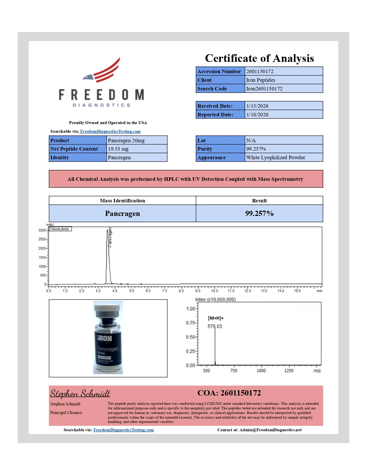
Molecular formula: C26H36N6O9
Molecular weight: 576.25 g/mol
Sequence: Lys-Glu-Asp-Trp
Other known titles: KEDW, DWa, amidate
Medical iron peptides
Observed Research Findings in Laboratory Models
In the information below, we have broken down the latest clinical and preclinical research on Pancragen’s potential as observed in various experimental models.
Pancragen and the Pancreas
The tetrapeptide Pancragen has been studied for its potential impact on pancreatic cells, particularly in relation to cell differentiation and the regulation of insulin and glucagon release, crucial elements in the pancreatic cells’ endocrine function. (1) Research posited that Pancragen might penetrate cellular membranes to interact with the nucleus and nucleolus, thereby possibly influencing the transcription of genes crucial for cellular differentiation within the pancreatic gland. Key differentiation factors such as Ptf1a, Pdx1, Pax6, Foxa2, Nkx2.2, and Pax4 are crucial for the proper functioning and differentiation of various pancreatic cell types. It is posited that Pancragen’s interaction may potentially up-regulate the expression of these factors, which may play a role in the cellular maturation process of both acinar and islet cells in the pancreatic gland. The experiments conducted on embryonic cultures of pancreatic acinar cells in this study suggested that Pancragen might significantly enhance the expression of Ptf1a and Pdx1 proteins, which are known to be crucial for the maturation of acinar and islet cells. The study suggested that Pancragen’s actions might be more pronounced in aged cultures, where an apparent decrease in the expression of these proteins is observed as a part of the cellular aging process. This potential up-regulation of key differentiation factors by Pancragen is posited to possibly lead to an increased differentiation of pancreatic cells, and, subsequently, may studied for associations with pancreatic cell signaling and differentiation. The researchers also posited that transcription factors that regulate differentiation of pancreatic cells are a pharmacological target for Pancragen, which allows considering it as applied in experimental models of pancreatic signaling and metabolic dysfunction.
Another research study using murine models to gauge the impact of Pancragen on the pancreas’s functional morphology, also yielded interesting findings. (2) Initially, when DM was induced in murine models, there was a noted decrease in insulin-producing B cells and an uptick in glucagon-producing A cells. Such changes point to disrupted pancreatic cell functionality. However, upon exposure to Pancragen, potentially encouraging shifts were observed. Murine models showed evident compensatory changes in pancreatic cells and tissue. Specifically, Pancragen seemed to bolster insulin production from B cells while tempering glucagon production from A cells. Additionally, the proliferative activity of certain cells and their apoptosis seemed to normalize, closely resembling control murine models.
Further research has also suggested that Pancragen might play a pivotal role in the modulation of various cellular markers and proteins associated with the vitality of pancreatic cells. When applied to aging pancreatic cells, there was an observed increase in the expression of matrix metalloproteinase MMP2 and MMP9, serotonin, glycoprotein CD79alpha, the antiapoptotic protein Mcl1, and proliferation markers PCNA and Ki67. Conversely, there was a decrease in the expression of the proapoptotic protein p53 in aged pancreatic cell cultures. From these observations, it can be posited that the tetrapeptide potentially holds the capability to activate the expression of signaling molecules that serve as markers of the functional activity of pancreatic cells. (3)
Pancragen and Metabolic Problems
Studies have delved into the potential of Pancragen on metabolic problems in aged test subjects. For example, in one study, the focus was mainly on carbohydrate metabolism and the potential role Pancragen may play in regulating it. (4) The introduction of Pancragen was observed to significantly lower fasting glucose levels during a standard glucose tolerance test, alongside a reduction in insulin concentrations and the insulin resistance index. This suggests a potential utility of Pancragen in addressing the disturbances in carbohydrate metabolism, especially given the observed persistence of its glucose-lowering action post-exposure. It is critical to note that the study was focused on august test subjects, and the scope of Pancragen’s potential may be contingent on several other variables, including the severity of insulin resistance and other factors. The researchers also commented that administration of the tetrapeptide Pancragen is a promising approach to the examined in preclinical models of insulin signaling and carbohydrate metabolism.
Another study aimed to investigate the potential impact of Pancragen on the endocrine function of the pancreatic cells and the metabolic status of test models. (5) Aged test models used in this research were used to understand the potential of Pancragen in addressing age-related dysfunctions in the overall metabolism as well as the pancreatic islet apparatus. In older test models, there was a noted reduced rate of glucose utilization compared to younger counterparts. Moreover, higher insulin and C-peptide peaks were observed 5 and 15 minutes post-glucose introduction. However, when Pancragen was introduced per test model daily over 10 days, there was a marked improvement in the glucose utilization rate. This intervention also seemed to studied for modulation of endocrine signaling pathways in experimental systems. Intriguingly, the actions of Pancragen lingered, with some aspects of metabolic status and pancreatic cell function remaining improved even three weeks after the cessation of the trial.
In a study examining the actions of Pancragen on endothelial function within the setting of chronic hyperglycemia using murine models, it was observed that the introduction of Pancragen may have potential role in examined in laboratory models of endothelial signaling and adhesion pathways. (6) Restoring these endothelial adhesive characteristics is crucial because, in models of metabolic problems, proper endothelial function plays a vital role in blood flow regulation and preventing complications such as atherosclerosis. Deficiencies or abnormalities in endothelial adhesion may increase susceptibility to vascular damage, making this restoration a key target for averting it.
Pancragen and Aging
Pancragen has been posited as a possible bioregulator for adjusting metabolic problems associated with aging. To understand Pancragen’s impact on aging, a study examined its potential on murine models across different age groups. (7) There was a noted decrease in certain aging biomarkers like caspase-3 and cathepsin B activities when Pancragen was introduced to the younger murine models. Interestingly, in the mature murine models, Pancragen application resulted in a significant reduction of TNF-α levels and an elevation in IGF-I, both of which are critical indicators related to aging and metabolic processes. The study also delved into a rapid experimental aging model by inducing diabetes mellitus in murine models. It was observed that Pancragen potentially normalized blood glucose levels, emphasizing its previously identified hypoglycemic properties. Furthermore, the presence of Pancragen might have suppressed certain apoptotic enzymatic components in the pancreatic cells, suggesting that its biological actions may be associated with both metabolic correction and antiapoptotic mechanisms. The actions of Pancragen on natural biological aging are potentially realized at the IGF-I level, known for its studied in aging pathway models for associations with IGF-I signaling and proteolytic regulation. Moreover, the tetrapeptide’s potential interaction with certain genes suggests that its influence might extend to the proteolytic processing level.
According to another trial, Pancragen has been posited to tissue-specifically influence gene expression in pancreatic cell cultures. (8) The study suggests that variations in the methylation patterns of the PDX1, PAX6, and NGN3 gene promoter regions in pancreatic cells may be associated with aging and could potentially be the cause behind the alterations in their expression levels. This implies that long-standing changes in gene expression upon aging might be driven by modifications in these promoter methylation patterns. However, the expression levels of the PAX4 and FOXA2 genes in pancreatic cells seem to deviate during aging and in response to Pancragen, even when the methylation patterns of the PAX4 gene remain stable. Curiously, the FOXA2 gene’s promoter region in pancreatic cells exhibited only a handful of methylated CpG sites, with their methylation levels being influenced by both cell culture aging and Pancragen. Yet, this did not correlate straightforwardly with changes in gene expression levels. This suggests that, despite the influence of Pancragen on certain gene methylation patterns, the exact relationship between methylation and gene expression may be intricate and possibly regulated by other, yet unidentified, mechanisms.
Pancragen peptide is available for research and laboratory purposes only.


Reviews
There are no reviews yet.